Retinal Vein Occlusions
What is a retinal vein occlusion?
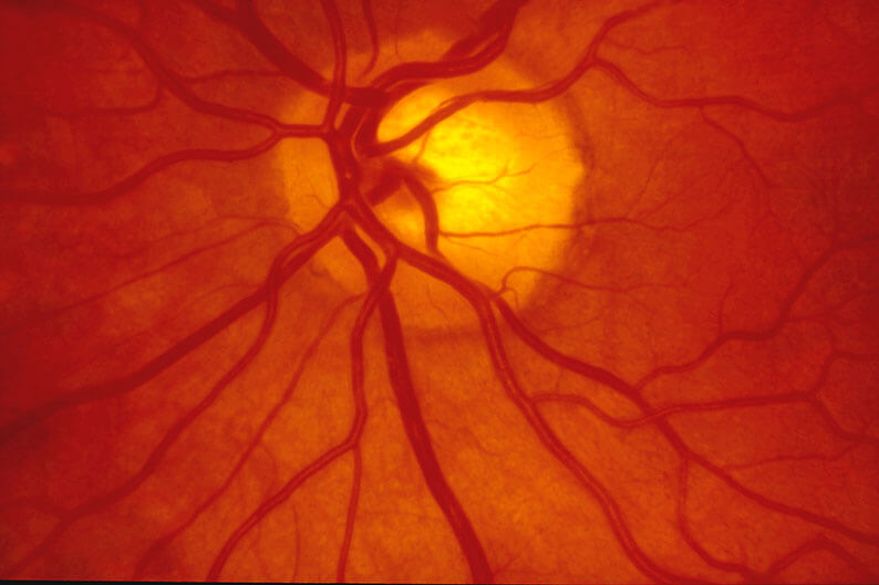
A retinal vein occlusion means that a vein in the retina of the eye has become blocked. The retina is the light sensing tissue at the back of the eye. The veins drain blood out of the retina and return it to the heart. Blockage or occlusion in the vein prevents adequate blood flow in the affected area. The walls of the blocked vein tend to leak blood and excess fluid into the retina. The retina in the area that is drained by that particular occluded vein then becomes swollen and functions poorly.
What are the types of retinal vein occlusion?
There are two types of retinal vein occlusion. In central retinal vein occlusion (CRVO), the main retinal vein is blocked. Blood flow is poor throughout the entire retina. The amount of visual loss varies, but may be severe. In branch retinal vein occlusion (BRVO), a smaller branch of the main retinal vein is blocked. Only the part of the retina drained by this branch vein is damaged. Vision loss varies but is not as severe as in CRVO. Retinal vein occlusions are more common in people who have high eye pressure, diabetes, age-related vascular (blood vessel) disease, or high blood pressure.
What are the symptoms of retinal vein occlusion?
Blurred vision is the main symptom of retinal vein occlusion. It occurs when the excess fluid leaking from the vein collects in the macula. The macula is the central area of the retina which is responsible for our central, detailed vision. If the macula swells with excess fluid (macular edema), vision becomes blurred.